


All Issues
X. fragariae and C. cladosporioides cause strawberry blossom blight
Publication Information
California Agriculture 53(4):26-28. https://doi.org/10.3733/ca.v053n04p26
Published July 01, 1999
PDF | Citation | Permissions
Abstract
Blossom blight was documented in some strawberry production fields in Watsonville in 1996 and 1997. Xanthomonas fragariae and Cladosporium cladosporioides were identified as the causal organisms. This is the first documentation of the two organisms causing blossom blight of strawberry in California. This is also the first report identifying C. cladosporioides as a pathogen of strawberry.
Full text
California strawberry production accounts for approximately 80% of the 1,500 million to 1,600 million pounds of strawberries produced annually in the United States. Of the fruit produced in California, more than 50% comes from the Central Coast, specifically the Salinas and Watsonville areas.
In the spring of 1996, a severe incidence of blossom blight occurred in some fruit production fields in the Watsonville area. In addition to blighting of entire flowers, the symptoms observed were watersoaked lesions on the lower surface of the calyx that appeared dark green under reflected light and translucent under transmitted light; necrotic calyces of seemingly healthy green and ripe fruits; water-soaking of the base of the calyx that extended into the pedicel; presence of green-gray sporulation on dead anthers; and presence of flower clusters with small and irregularly shaped fruits. Most of the affected plants showed veinal watersoaking and angular lesions on the lower leaf surfaces, symptoms typical of angular leaf spot caused by Xanthomonas fragariae. Blossom blight was observed again in 1997, but fewer fields were affected and the disease was less severe than in the previous year. In this paper, we report the association of X. fragariae and Cladosporium cladosporioides with strawberry blossom blight in California.
Identification techniques
Microscopic examination and isolation.
We collected diseased flowers from several fields for microscopic observation and isolation of possible causal agents. Pieces of tissue from diseased flower parts were either macerated in a few drops of sterile distilled water and streaked on 2.3% nutrient agar, 0.5% glucose and 0.1% yeast extract (NDYA) and acidified potato-dextrose agar (APDA) or plated directly on these media, before or after surface sterilization. Surface sterilization consisted of dipping tissue pieces in 0.5 % NaOCl for 30 seconds and rinsing twice with sterile distilled water. We made successive transfers onto NDYA or APDA to obtain pure cultures of isolated bacteria and fungi, respectively.
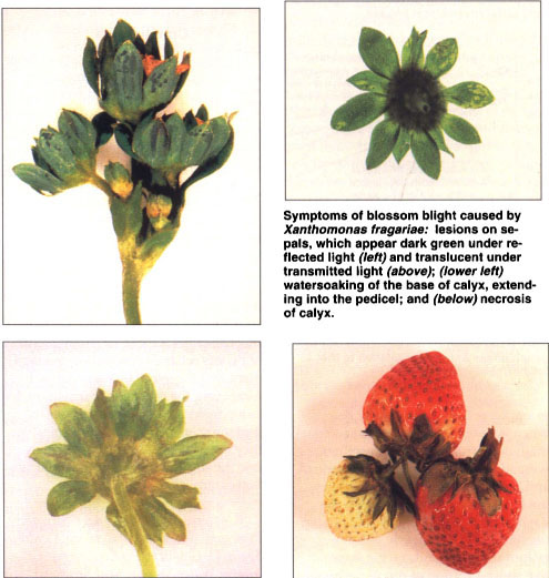
Symptoms of blossom blight caused by Xanthomonas fragariae: lesions on sepals, which appear dark green under reflected light left and translucent under transmitted light above; lower left watersoaking of the base of calyx, extending into the pedical; and below necrosis of calyx.
Pathogenicity tests.
We used greenhouse-grown 'Selva' strawberry plants to determine pathogenicity of the bacterial and fungal isolates. Inoculation with the bacteria was done by airbrush spraying (applied at a pressure of 1 kg/cm2) the flowers and lower leaf surfaces with aqueous cell suspensions containing approximately 108 colony-forming units/ml (A600 = 0.1) prepared from 48-hour-old cultures of the bacteria. Inoculation with the fungal isolate was done by spraying spore suspensions containing approximately 106 conidia/ml onto the flowers until runoff, using a plastic spray bottle. Plants were inoculated with either the bacteria, fungi or both. We covered inoculated and control (sprayed with sterile distilled water) plants with plastic bags to maintain a high relative humidity and incubated them in the laboratory (24±2°C; about 75±4°F) and greenhouse (24±4°C; about 75±7°F) for 2 to 5 days. To complete Koch's postulates, isolations were made from symptomatic inoculated plant parts.
Polymerase chain reaction (PCR).
Genomic fingerprinting, based on PCR using specific primers for repetitive DNA sequences, was also used to identify selected bacterial strains isolated from the flowers. Two methods were used: enterobacterial repetitive intergenic consensus ERIC-PCR and repetitive extragenic palindromic REP-PCR. The analyses were performed as described previously for X. fragariae (Roberts et al. 1996; Opgenorth et al. 1996).
Bacteria and fungus isolated
Microscopic observation.
We observed bacterial streaming in 100% of sepals and peduncles that showed watersoaking; in 25% to 35% of necrotic sepals, anthers and pistils; and in about 5% of necrotic petals.
Isolation.
Yellow bacterial colonies were consistently isolated from water-soaked or necrotic lesions on calyces and pedicels. These colonies were entire, circular, raised, glistening, mucoid and slow growing — characteristics typical of X. fragariae on NDYA. In addition to the yellow bacteria, a fungus was frequently isolated from infected anthers, sepals, petals and pistils. On PDA, the fungus appeared as velvet colonies colored olivaceous-green to olivaceous-brown with conidiophores that were branched both apically and laterally. Conidia were lemon-shaped and usually smooth, but sometimes textured. This fungus was identified as C. cladosporioides.
Pathogenicity tests.
Leaves of plants inoculated with X. fragariae developed small watersoaked lesions within 5 to 7 days of incubation. Symptoms on the calyx appeared within 7 to 10 days as minute water-soaked lesions that later coalesced and turned dark brown and necrotic. Watersoaked lesions on the pedicel, which usually started at the calyx base, became visible after 10 days. Occasionally, infected pedicels collapsed, leading to blighting of the attached flower or developing fruit. Lesions on leaves and sepals appeared dark green under reflected light and translucent under transmitted light. These were indistinguishable from lesions associated with angular leaf spot caused by X. fragariae. Blossoms inoculated with C. cladosporioides showed discoloration within 5 to 7 days. The anthers turned black, were covered with green-gray sporulation and eventually became necrotic. Petals, sepals, entire flowers or developing fruits also became necrotic. Some flower clusters produced small and irregularly shaped fruits. Plants inoculated simultaneously with both organisms produced all of the symptoms described above. Noninoculated control plants remained asymptomatic. X. fragariae and C. cladosporioides were reisolated from inoculated symptomatic plants, not from control plants.
PCR.
ERIC- and REP-PCR banding patterns of 15 bacterial strains isolated from strawberry flowers and X. fragariae reference strain 33239 were identical. In addition, these PCR-based genomic fingerprints were identical to 12 X. fragariae strains previously isolated from strawberry leaves in California and one strain isolated from Florida.
New fungus causes blight
Xanthomonas fragariae and Cladosporium cladosporioides were isolated from strawberry flowers with symptoms of blight. Blossoms inoculated with these organisms developed symptoms distinct from each other. The most prominent visible symptom caused by the bacterium X. fragariae prior to blighting of the flower was watersoaking of the pedicel that usually started at the calyx end. Infection by the fungus C. cladosporioides is characterized by necrosis of flower parts or the entire flower, presence of green-gray sporulation on dead anthers and production of small and malformed or misshapen fruits. The production of malformed fruits is probably due to poor pollination resulting from anther or pistil infection. Large numbers of unfertilized ovules lead to production of undeveloped or misshapen fruits in strawberries (Connors 1972).
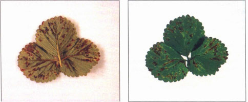
Strawberry leaflet showing typical symptoms of angular leaf spot caused by Xanthomonas fragariae: veinal watersoaking and angular lesions on the lower surface, which appear dark green under reflected light (left) and translucent under transmitted light (right).
In 1996, all affected fields showed disease symptoms caused by both X. fragariae and C. cladosporioides. In 1997, some fields were affected by both organisms, while others showed symptoms caused only by one or the other organism. Although infection by X. fragariae seemed to aggravate the effect of C. cladosporioides, and vice versa, each organism was capable of causing blossom blight independently. In the fields observed in this study, C. cladosporioides was the predominant cause of blossom blight.
X. fragariae can cause occasional fruit loss due to pedicel collapse, but this is considered of minor economic importance in California strawberry production fields. More significant is calyx infection, which renders the fruit unattractive and therefore reduces the market value of the crop.
Although we did not estimate yield losses due to infection by C. cladosporioides, two growers lost strawberry yield completely for one entire harvest cycle in 1996. Environmental conditions favorable for disease development could result in this disease complex being a major factor in limiting production. Until now blossom blight of strawberry in California has been mostly attributed to Botrytis cinerea. This is the first report that C. cladosporioides can cause blossom blight in strawberry. This is also the first report that this organism is a pathogen of strawberry.
Blossom blight was first observed in June 1996 following a relatively heavy rainfall on May 31. The conditions that led to severe incidence of the disease are not clear. Precipitation was almost 4 inches in May 1996 and less than 1 inch in May 1995 and 1997. Leaves stayed wet for longer duration in 1996 than in 1995 or 1997. The daily leaf wetness period in May ranged from 10 to 24 hours (average = 16 hours) in 1996,0 to 20 hours (average = 11 hours) in 1995, and 8 to 22 hours (average = 14 hours) in 1997. A combination of iprodione (Kocide 101 at 2 lb/ac), fixed copper (Rovral 50W at 1.5 lb/ac) and methomyl (Lannate at 1 lb/ ac) spray apparently controlled the malady, but it is not clear whether all or one particular chemical was responsible for the results obtained. It is also possible that the warmer and drier weather in July helped suppress the disease.

Symptoms of blossom blight induced by Cladosporium cladosporioides: (upper left) presence of green-gray sporulation on anthers; (upper right) necrosis of flower; and (above) presence of flower clusters with small and abnormally shaped fruits.
The effect of weather on the incidence and severity of blossom blight caused by X. fragariae and C. cladosporioides, and the development of effective control measures need further investigation
X. fragariae and C. cladosporioides cause strawberry blossom blight